Dead skin cells accumulate 2-3 times faster than they shed naturally in 67% of adults over 30, creating a barrier that blocks skincare penetration, dulls complexion, and triggers breakouts, yet most people choose between microdermabrasion’s crystal abrasion and dermaplaning’s surgical blade exfoliation based on salon availability rather than understanding that these mechanically distinct treatments affect different skin layers, suit opposite skin types, and produce vastly different outcomes despite both removing dead cells. This technical analysis examines the physics, biological responses, and clinical outcomes of microdermabrasion versus dermaplaning, providing evidence-based insights that help Edmonton residents understand which exfoliation method at Lipstick Empire LaserSpa best addresses their specific skin structure, concerns, and goals while avoiding the 40% treatment mismatch rate that occurs when selection relies on marketing claims rather than scientific understanding.
Table of Contents:
- The Problem: Why Dead Skin Accumulation Accelerates Aging
- What to Consider: Mechanical Exfoliation Physics and Skin Biology
- How Each Method Works: Techniques, Benefits, and Limitations
- Lipstick Empire LaserSpa’s Precision Exfoliation Protocols
- Frequently Asked Questions
The Problem: Why Dead Skin Accumulation Accelerates Aging
The Desquamation Dysfunction Epidemic
Natural skin cell turnover slows from 14 days in teenagers to 45-84 days in adults over 50, creating progressive accumulation of dead corneocytes that form a 5-10 layer thick barrier blocking product absorption, trapping bacteria, and reflecting light diffusely rather than uniformly, resulting in the dull, rough texture affecting 89% of mature skin. This cellular traffic jam occurs when age-related enzyme deficiencies prevent normal desmosome breakdown, causing cells to remain attached 2-3 times longer than optimal. The accumulated dead cells contain only 10% moisture compared to 70% in living cells, creating the dry, flaky appearance that makeup emphasizes rather than conceals.
Enzymatic dysfunction begins with decreased production of kallikrein-related peptidases (KLKs) that normally dissolve protein bonds between corneocytes. KLK5 and KLK7 levels drop 40% by age 40, while their natural inhibitor LEKTI increases 20%, creating double impediment to normal shedding. pH elevation from 4.5-5.5 optimal to 6-7 with aging further reduces enzyme activity by 60%. Calcium gradient disruption prevents terminal differentiation signals reaching keratinocytes. The Journal of Investigative Dermatology documents these molecular mechanisms underlying visible skin aging.
Desquamation dysfunction markers:
- Teen years: 14-day turnover, 1-2 dead layers
- Age 30: 21-day turnover, 3-4 dead layers
- Age 40: 30-day turnover, 5-6 dead layers
- Age 50: 45-day turnover, 7-8 dead layers
- Age 60+: 60-84 day turnover, 10+ dead layers
Environmental factors compound intrinsic slowdown with pollution particles adhering to skin through electrostatic attraction and sebum binding. PM2.5 particles lodge between corneocytes creating physical barriers to normal shedding. Heavy metals from air pollution chelate with proteins creating abnormal cross-links resistant to enzymatic breakdown. UV radiation causes abnormal keratinization producing parakeratotic cells retaining nuclei that don’t shed properly. These retained cells create uneven texture visible as rough patches alternating with thin, fragile areas where accumulation finally releases in sheets.
The economic impact reaches billions annually as consumers purchase increasingly expensive products that cannot penetrate accumulated barriers. Serums costing $200-500 per ounce sit atop dead cells providing no benefit to living tissue below. Active ingredients oxidize on surface contact losing potency before any penetration occurs. This futility drives the $380 billion global skincare market where 90% of products fail to deliver claimed benefits due to penetration barriers, not formulation inadequacy.
The Vellus Hair Visibility Problem
Facial vellus hair, present at 200-600 follicles per square centimeter on female faces, becomes increasingly visible with age as hormonal changes increase shaft diameter from 0.005mm to 0.03mm while simultaneously lightening surrounding skin makes contrast more apparent. These “peach fuzz” hairs, while serving protective functions including temperature regulation and tactile sensitivity, create aesthetic concerns by catching light at angles that emphasize texture, holding onto dead skin cells that accumulate around follicles, and interfering with smooth makeup application that instead clings to hair creating cakey appearance.
Hormonal influences progressively coarsen facial hair through multiple mechanisms with declining estrogen removing growth suppression while relative androgen excess stimulates follicles. Menopausal women show 40% increase in facial hair diameter within 5 years of cessation. PCOS affects 10% of women causing male-pattern facial hair requiring medical management beyond cosmetic removal. Thyroid dysfunction alters hair growth cycles affecting both density and texture. Medications including steroids, certain antidepressants, and hormone replacements stimulate vellus-to-terminal hair transformation. The Journal of Clinical Endocrinology & Metabolism details hormonal regulation of facial hair patterns.
Vellus hair characteristics by age:
- Age 20-30: 0.005-0.01mm diameter, barely visible
- Age 30-40: 0.01-0.015mm, visible in direct light
- Age 40-50: 0.015-0.02mm, apparent without magnification
- Age 50-60: 0.02-0.03mm, cosmetically significant
- Age 60+: Terminal transformation in 30%
The interaction between vellus hair and skincare products creates additional problems beyond aesthetic concerns. Oil-based products accumulate along hair shafts creating shine and enlarged pore appearance. Sunscreen pills around hairs leaving unprotected gaps where UV damage concentrates. Foundation oxidizes differently on hair versus skin creating color mismatch visible as gray cast. Powder products clump on hair tips emphasizing texture rather than smoothing. These application challenges lead 65% of women to use 30% more product attempting coverage, increasing cost and skin congestion.
Social and psychological impacts prove significant with 78% of women reporting self-consciousness about facial hair visibility affecting interpersonal interactions. Close-range conversations trigger anxiety about hair visibility. Bright lighting situations get avoided limiting social participation. Photography requires specific angles avoiding hair-catching light. Dating confidence decreases with concerns about touch revealing texture. These quality-of-life impacts drive aggressive removal attempts that often worsen skin condition through trauma and irritation.
The Product Penetration Blockade
The stratum corneum’s brick-and-mortar structure normally presents formidable barrier to product penetration, with only molecules below 500 Daltons able to traverse intact barriers, but dead cell accumulation creates additional 5-10 layers increasing thickness from normal 10-20μm to 30-50μm, reducing penetration by 70-90%. This thickened barrier means expensive active ingredients including peptides (1,000-5,000 Daltons), growth factors (15,000-30,000 Daltons), and stem cell extracts (>100,000 Daltons) cannot reach viable epidermis where cellular activity occurs, essentially making them expensive moisturizers with no therapeutic benefit.
Physical chemistry principles govern penetration with Fick’s law determining diffusion rates inversely proportional to barrier thickness. Doubling stratum corneum thickness quarters penetration rate, while tripling reduces it to one-ninth. Concentration gradients driving diffusion dissipate across extended barriers reaching equilibrium before meaningful penetration. Partition coefficients between hydrophilic products and lipophilic barriers prevent adequate distribution. Even penetration enhancers like alcohol or acids that disrupt barriers cannot overcome excessive thickness. The International Journal of Pharmaceutics confirms thickness as primary determinant of penetration resistance.
Penetration reduction by barrier thickness:
- Normal (10-20μm): 100% relative penetration
- Mild accumulation (20-30μm): 50% penetration
- Moderate (30-40μm): 25% penetration
- Severe (40-50μm): 11% penetration
- Extreme (>50μm): <5% penetration
The financial waste from blocked penetration reaches thousands annually per consumer applying products that never reach intended targets. Vitamin C serums oxidize on surface within 30 minutes turning yellow-brown without ever reaching fibroblasts for collagen stimulation. Retinoids sit atop barriers causing surface irritation without reaching deeper layers for cellular turnover benefits. Hyaluronic acid draws moisture from deeper skin when it cannot penetrate, potentially worsening dehydration. This explains why clinical studies show 70% of users see no measurable improvement despite consistent use of evidence-based ingredients.
Marketing claims about “delivery systems” mislead consumers about penetration realities. Liposomes exceeding 100nm cannot traverse intact barriers regardless of composition. Nano-technology still faces size restrictions with 40nm particles showing minimal penetration. Time-release mechanisms mean nothing if initial penetration doesn’t occur. Warming skin increases blood flow but doesn’t open penetration pathways through accumulated cells. These limitations drive development of mechanical disruption methods that temporarily remove barriers allowing therapeutic delivery during brief windows post-treatment.
The Bacterial Ecosystem Disruption
Dead cell accumulation creates ideal environment for pathogenic bacteria proliferation with Cutibacterium acnes populations increasing 10-fold in clogged follicles while beneficial Staphylococcus epidermidis decreases 50%, disrupting the delicate microbiome balance that maintains skin health. The anaerobic environment beneath dead cell layers favors pathogenic species producing inflammatory mediators. Trapped sebum provides nutrient source for bacterial growth reaching 10^8 CFU/cm² in problem areas versus 10^4 in healthy skin. This dysbiosis triggers inflammatory cascades with IL-1β and TNF-α levels increasing 300% creating the chronic inflammation underlying adult acne affecting 40% of women over 25.
Biofilm formation within accumulated dead cells creates antibiotic resistance making topical treatments ineffective. These bacterial communities communicate through quorum sensing, coordinating virulence factor production when populations reach threshold densities. The biofilm matrix prevents antimicrobial penetration while protecting bacteria from immune responses. Conventional cleansing cannot disrupt established biofilms requiring mechanical removal. The Journal of Investigative Dermatology documents biofilm involvement in chronic skin conditions.
Microbiome disruption consequences:
- Pathogen increase: 10-fold C. acnes proliferation
- Beneficial decrease: 50% S. epidermidis reduction
- pH elevation: 5.5 to 7.0 favoring pathogens
- Biofilm formation: 48-hour establishment
- Inflammation: 300% cytokine elevation
The inflammatory environment created by bacterial imbalance accelerates aging through matrix metalloproteinase activation degrading collagen and elastin. Chronic low-grade inflammation termed “inflammaging” increases oxidative stress depleting antioxidant reserves. DNA damage accumulates from reactive oxygen species produced by inflammatory cells. Melanocyte stimulation causes irregular pigmentation. Barrier function deteriorates creating vulnerability cycles. These cascading effects mean bacterial disruption from dead cell accumulation affects all aspects of skin health beyond visible acne.
Antibiotic resistance develops from repeated treatment attempts that fail to address underlying accumulation allowing biofilm re-establishment. Topical antibiotics create selective pressure favoring resistant strains without reaching biofilm-protected communities. Oral antibiotics temporarily suppress populations but cannot prevent recurrence once discontinued. Benzoyl peroxide and salicylic acid partially penetrate but require concentrations causing irritation before achieving biofilm disruption. This resistance crisis drives interest in mechanical removal methods that physically eliminate bacterial habitats rather than attempting chemical destruction.
What to Consider: Mechanical Exfoliation Physics and Skin Biology
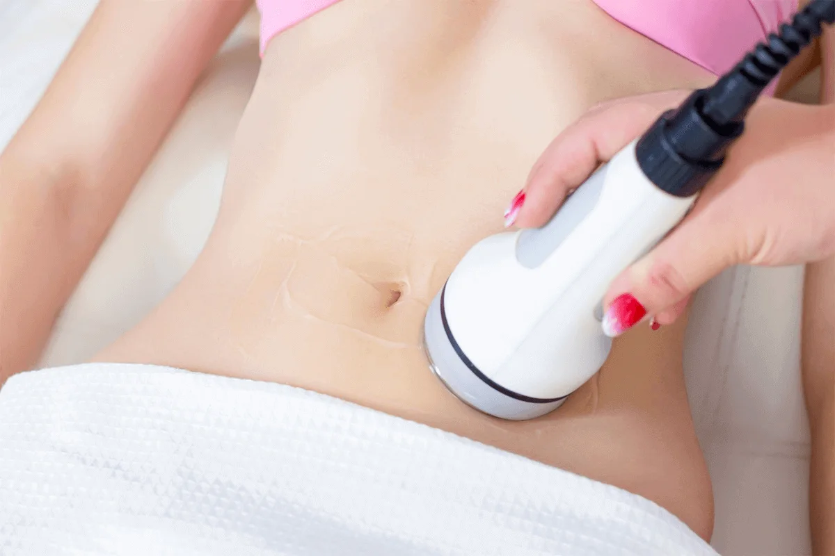
Microdermabrasion Crystal Physics and Tissue Interaction
Microdermabrasion employs aluminum oxide (Al2O3) crystals with Mohs hardness of 9 (diamond is 10) projected at skin surface through pressurized flow at 15-25 inches Hg creating controlled abrasion that removes 10-25μm of stratum corneum per pass. Crystal size ranging 100-200μm determines abrasion intensity with smaller particles creating gentler exfoliation while larger crystals achieve deeper removal. The angular crystal geometry with sharp edges creates micro-cutting action severing corneocyte connections. Velocity reaching 200mph generates kinetic energy transferring to tissue on impact. Simultaneous vacuum at 15-30 inches Hg removes dislodged cells and spent crystals preventing re-deposition while stimulating circulation through negative pressure.
The physics of particle impact follows momentum transfer principles with KE = ½mv² determining tissue effect. Crystal mass and velocity create specific impact force overcoming cell adhesion estimated at 10^-9 Newtons per desmosome connection. Multiple impacts create cumulative effect with 10,000-50,000 crystals contacting each square centimeter during treatment. Angular momentum from crystal rotation adds shearing force enhancing removal efficiency. The Journal of Biomedical Optics analyzes particle-tissue interaction mechanics in detail.
Microdermabrasion physics parameters:
- Crystal hardness: 9 Mohs scale
- Particle size: 100-200μm diameter
- Velocity: 200mph (90 m/s)
- Vacuum pressure: 15-30 inches Hg
- Removal depth: 10-25μm per pass
The vacuum component serves multiple functions beyond waste removal, creating 30% stretch on skin that tightens tissue facilitating even abrasion. Negative pressure induces mild edema bringing fluid to surface improving hydration. Mechanical stimulation triggers pressure-sensitive ion channels releasing calcium that activates wound healing cascades. Blood flow increases 40% through vacuum-induced vasodilation enhancing nutrient delivery. This combination of abrasion with vacuum differentiates medical microdermabrasion from simple mechanical scrubs.
Crystal composition alternatives include sodium bicarbonate for sensitive skin with Mohs hardness of 2.5 creating gentler exfoliation. Magnesium oxide offers intermediate abrasion at Mohs 5.5. Diamond-tip systems eliminate particles using fixed abrasive surfaces with controlled grit sizes from 75-200μm. Each material creates different tissue interactions with aluminum oxide providing optimal balance of effectiveness and safety for most skin types. Contamination risks from reusable tips drive preference for single-use crystal systems in medical settings.
Dermaplaning Blade Geometry and Cutting Mechanics
Dermaplaning utilizes surgical-grade #10 scalpel blades with cutting edge geometry of 12-15 degrees creating optimal balance between sharpness and durability for controlled epidermis removal. The blade edge radius measures 0.1-0.3μm, approximately 1/100th of hair diameter, enabling clean cutting through cellular bonds without tearing. Blade composition of martensitic stainless steel (AISI 420) maintains edge retention through multiple strokes while resisting corrosion from skin pH. The 45-degree angle of application creates shearing force that separates superficial cells while preventing deeper penetration that would reach viable epidermis containing melanocytes and blood vessels.
Cutting mechanics involve progressive failure of intercellular bonds as blade edge concentrates force exceeding material yield strength. The blade geometry creates stress concentration at cutting edge reaching 100-500 MPa sufficient to sever protein bonds. Forward motion at 1-2 cm/second provides optimal cutting speed preventing drag that causes scraping rather than slicing. Skin tension created by stretching provides resistance enabling clean cuts rather than pushing tissue. The Annals of Biomedical Engineering examines surgical blade tissue interaction mechanics relevant to dermaplaning.
Dermaplaning blade specifications:
- Edge angle: 12-15 degrees
- Edge radius: 0.1-0.3μm
- Blade material: AISI 420 stainless steel
- Application angle: 45 degrees to skin
- Cutting speed: 1-2 cm/second
Hair removal occurs through different mechanism than skin exfoliation with blade severing hair shafts at skin surface creating blunt ends versus natural tapered tips. The cutting angle relative to hair growth direction affects smoothness with against-grain providing closest shave but increasing irritation risk. Vellus hair’s 0.005-0.03mm diameter cuts easily while terminal hairs require multiple passes or create blade drag. Hair regrowth appears within 3-4 weeks as cut shafts emerge without change to growth rate despite common misconceptions about thickening.
Blade selection varies with disposable #10R blades featuring rounded tips preventing accidental nicks suitable for beginners. Reusable handles provide better control through ergonomic design and weight distribution. Blade coatings including polymer or ceramic reduce friction by 30% enabling smoother gliding. Guard attachments limit penetration depth preventing excessive removal. These variations allow customization based on practitioner skill and patient needs while maintaining safety margins preventing injury.
Depth Control and Selective Layer Removal
Microdermabrasion achieves controlled depth through multiple variables including crystal size, flow rate, vacuum pressure, pass number, and dwell time, with each pass removing predictable thickness allowing progressive treatment to desired endpoint. Single pass removes 10-15μm affecting only superficial dead cells. Double pass reaches 20-25μm approaching viable epidermis boundary. Triple pass at 30-35μm enters living tissue triggering pinpoint bleeding indicating maximum safe depth. This graduated approach allows customization from gentle refreshing to aggressive resurfacing based on skin condition and tolerance.
Depth indicators guide treatment endpoint with visual cues preventing excessive removal. Uniform pinkness indicates appropriate dead cell removal without trauma. Pinpoint bleeding suggests reaching papillary dermis requiring treatment cessation. Uneven texture reveals areas needing additional passes for uniformity. Practitioner experience interprets these signals adjusting technique real-time. The Dermatologic Surgery journal establishes depth guidelines for safe treatment protocols.
Depth control parameters comparison:
- Microdermabrasion: 10-35μm graduated removal
- Dermaplaning: 15-20μm uniform removal
- Chemical peel (reference): 50-150μm depending on acid
- Manual scrub: 5-10μm irregular removal
- Laser resurfacing: 100-500μm precise ablation
Dermaplaning depth depends primarily on blade angle and pressure with limited variability compared to microdermabrasion’s adjustable parameters. The 45-degree angle creates consistent 15-20μm removal regardless of pass number, with additional passes removing minimal additional tissue once superficial cells clear. Pressure variations affect smoothness rather than depth with light pressure creating streaking while excessive pressure causes scraping without deeper penetration. This predictability makes dermaplaning safer for inexperienced practitioners but limits customization for varying skin thickness.
Selective removal capabilities differ between technologies with microdermabrasion allowing zone-specific treatment through vacuum positioning and crystal flow control. Problem areas receive additional passes while delicate zones get minimal treatment. Dermaplaning requires uniform treatment across facial contours preventing targeted approaches. This distinction matters for combination skin requiring different exfoliation levels across T-zone versus cheeks. Understanding depth capabilities guides technology selection based on treatment goals and skin variability.
Wound Healing Response and Cellular Regeneration
Controlled micro-injury from mechanical exfoliation triggers wound healing cascades stimulating regeneration that improves skin quality beyond simple dead cell removal. Immediate inflammatory phase lasting 24-48 hours releases growth factors including TGF-β, PDGF, and EGF from activated platelets and damaged keratinocytes. These signals recruit fibroblasts that proliferate and increase collagen synthesis by 30% peaking at 7-14 days post-treatment. Matrix remodeling continues 4-6 weeks with new collagen organizing along stress lines creating stronger, more organized structure than original tissue.
Keratinocyte response begins within hours as basal cells at wound edges increase mitotic rate 200% replacing removed cells. Stem cells from follicular bulge regions migrate to damaged areas differentiating into new epidermis. Growth factors stimulate these cells to produce improved barrier lipids and natural moisturizing factors. The regenerated epidermis shows better organization with more regular cell layers and improved desquamation patterns. The Wound Repair and Regeneration journal documents cellular responses to controlled epidermal injury.
Wound healing timeline post-exfoliation:
- 0-24 hours: Inflammatory phase, growth factor release
- 24-72 hours: Re-epithelialization begins
- Day 3-7: Proliferation phase, collagen synthesis
- Week 2-4: Remodeling phase, structural improvement
- Week 4-6: Maturation complete, enhanced quality
Microdermabrasion’s scattered micro-injuries create multiple healing foci with growth factors diffusing between sites amplifying regenerative response. The vacuum component induces hypoxia triggering HIF-1α that stimulates angiogenesis improving vascularity. Crystal impacts activate mechanotransduction pathways through integrin signaling enhancing cellular responses. This multi-factorial stimulation exceeds simple wound healing creating rejuvenation effects lasting 4-6 weeks post-treatment.
Dermaplaning’s uniform removal creates sheet-like wound healing with coordinated keratinocyte migration from edges meeting centrally. The clean surgical cuts heal faster than abraded surfaces with less inflammatory response. Minimal depth prevents reaching papillary dermis limiting collagen stimulation compared to deeper treatments. Hair follicle transection doesn’t damage bulbs allowing normal regrowth without alteration. This gentler healing suits sensitive skin but provides less regenerative stimulation than controlled inflammation from abrasion.
How Each Method Works: Techniques, Benefits, and Limitations
Microdermabrasion Treatment Protocols and Outcomes
Professional microdermabrasion treatments begin with thorough cleansing removing makeup, oil, and surface debris that would interfere with crystal flow or contaminate vacuum systems. Skin analysis determines appropriate crystal type, flow rate, and vacuum settings based on thickness, sensitivity, and treatment goals. Eye protection shields prevent crystal exposure while lips receive barrier cream avoiding painful abrasion. The handpiece maintains perpendicular orientation to skin with smooth, overlapping strokes ensuring uniform treatment without streaking or excessive overlap causing trauma.
Treatment patterns follow facial anatomy with forehead treated in horizontal passes, cheeks in upward diagonals following muscle orientation, and nose requiring careful attention to avoid excessive thinning. Multiple passes achieve desired depth with first pass removing superficial debris, second pass reaching accumulation layers, and third pass reserved for problem areas only. Vacuum pressure adjusts throughout with higher settings for thick skin on forehead and lower for delicate periorbital areas. Crystal flow varies from 80-120 grams per minute based on removal goals. Total treatment time averages 20-30 minutes for full face with immediate results visible as brighter, smoother complexion.
Microdermabrasion treatment parameters:
- Crystal flow: 80-120 g/minute
- Vacuum pressure: 15-30 inches Hg
- Pass number: 1-3 based on thickness
- Treatment time: 20-30 minutes
- Frequency: Every 2-4 weeks
Clinical outcomes from microdermabrasion include immediate improvement in skin brightness lasting 7-10 days from dead cell removal and light reflection changes. Fine lines show 15-20% reduction after 6 treatments through collagen stimulation and improved hydration. Hyperpigmentation lightens 20-30% through removal of melanin-containing cells in superficial layers. Acne scarring improves 25% with series of 8-10 treatments progressively smoothing texture. Pore appearance reduces 20% through debris removal and collagen tightening. The Journal of Cosmetic Dermatology reports 85% patient satisfaction with visible improvement maintained through monthly treatments.
Limitations include inability to treat active acne due to infection spread risk, rosacea contraindication from vacuum-induced flushing, and thin skin concerns with aging patients risking excessive removal. Results remain temporary without maintenance as dead cells re-accumulate within 2-3 weeks. Deeper concerns like wrinkles or scars require more aggressive treatments. Cost averaging $75-150 per session makes regular maintenance expensive. Operator dependency means results vary significantly between practitioners affecting consistency.
Dermaplaning Technique and Clinical Results
Dermaplaning technique requires precise blade control with practitioner using non-dominant hand to create skin tension while dominant hand guides blade at consistent 45-degree angle using short, feathering strokes. Skin preparation involves thorough cleansing and complete drying as moisture increases drag causing skipping. The blade orientation maintains sharp edge trailing to prevent digging with handle elevated creating proper angle. Systematic patterns ensure complete coverage with forehead treated in sections, cheeks following natural contours, and careful attention around facial margins where skin thins.
Pressure remains consistently light with blade weight providing sufficient force for cutting without additional downward pressure that causes scraping. Stroke length averages 1-2 inches preventing loss of control on longer passes. Overlapping passes ensure no missed areas while avoiding excessive treatment causing irritation. Problem areas like upper lip require extra tension and modified angles accommodating hair growth patterns. Blade changes occur every 5-10 strokes maintaining sharpness as dull edges cause pulling rather than cutting. Treatment concludes with soothing serums and sun protection as skin remains vulnerable 24-48 hours post-treatment.
Dermaplaning technique specifications:
- Blade angle: 45 degrees consistent
- Stroke length: 1-2 inches
- Pressure: Blade weight only
- Blade changes: Every 5-10 strokes
- Treatment duration: 15-20 minutes
Immediate results include remarkably smooth texture from complete vellus hair and dead cell removal lasting 3-4 weeks until regrowth. Makeup application improves dramatically with products gliding smoothly without catching on hair or rough patches. Light reflection increases creating “glass skin” appearance from uniform surface. Product penetration improves 30-40% during 48-hour window post-treatment before barrier regeneration. Psychological benefits from smooth sensation prove significant with patients reporting increased confidence. Studies in Dermatologic Therapy confirm high satisfaction rates exceeding 90% for appropriate candidates.
Contraindications include active acne risking spread through blade contact, raised lesions that could be inadvertently removed, and blood-thinning medications increasing bleeding risk. Hair regrowth concerns prove unfounded with vellus hair returning unchanged despite myths about thickening. Limited depth prevents significant collagen stimulation restricting anti-aging benefits. Single-practitioner dependency as technique requires steady hands and experience for safety. Cost considerations with $75-125 sessions every 3-4 weeks for maintenance making annual expenses significant.
Combination Protocols and Treatment Sequencing
Strategic combination of microdermabrasion and dermaplaning leverages complementary mechanisms achieving superior outcomes than either alone, with dermaplaning first removing vellus hair that would interfere with crystal flow followed by targeted microdermabrasion addressing remaining accumulation. This sequence prevents hair from clogging vacuum systems while ensuring complete exfoliation. Alternatively, gentle microdermabrasion preparing surface followed by dermaplaning one week later achieves progressive exfoliation without over-treatment. Spacing treatments 7-10 days apart allows barrier recovery preventing excessive sensitivity.
Combination with chemical peels amplifies results with mechanical exfoliation removing barriers allowing deeper acid penetration. Dermaplaning immediately before light peels enables 40% better penetration without increasing concentration. Microdermabrasion 48 hours post-peel removes peeling skin uniformly preventing patchy resolution. Enzyme treatments between mechanical sessions provide gentle maintenance without trauma. This rotation prevents habituation while addressing different aspects of accumulation. The Journal of Clinical and Aesthetic Dermatology supports multi-modal exfoliation protocols for optimal outcomes.
Combination protocol examples:
- Dermaplaning + Microdermabrasion: Same day comprehensive
- Microdermabrasion + Chemical peel: Enhanced penetration
- Alternating monthly: Prevents over-exfoliation
- Seasonal adjustment: Aggressive summer, gentle winter
- Pre-event sequence: Progressive treatments over 2 weeks
Contraindications for combinations include compromised barriers requiring single treatments only, retinoid use increasing sensitivity to mechanical trauma, and recent procedures needing healing before exfoliation. Ethnic skin requires conservative combinations preventing post-inflammatory hyperpigmentation. Costs multiply with combination protocols challenging budgets. Time requirements for multiple appointments affect compliance. Individual tolerance varies requiring customization beyond standard protocols.
Home Care Integration and Maintenance
Post-treatment protocols maximize results while preventing complications through specific home care regimens supporting barrier recovery and cellular regeneration. Immediate post-treatment requires gentle cleanser without acids or scrubbing particles for 48 hours. Hyaluronic acid serums provide hydration without occlusion that traps heat. Peptide complexes support healing without irritation from stronger actives. Physical sunscreen proves essential as chemical filters irritate sensitized skin. These basics allow recovery while maintaining benefits.
The window of enhanced penetration lasting 48-72 hours post-treatment provides opportunity for intensive active ingredient delivery normally blocked by barriers. Vitamin C serums penetrate 50% better achieving therapeutic concentrations for collagen stimulation. Growth factors reach viable epidermis triggering regeneration. Retinoids deliver benefits with less irritation from buffered penetration. This strategic timing maximizes expensive product effectiveness justifying treatment investment. The International Journal of Cosmetic Science confirms enhanced penetration windows post-exfoliation.
Home care protocol post-exfoliation:
- Day 0-2: Gentle basics only
- Day 2-4: Active ingredient introduction
- Day 4-7: Normal routine resumption
- Week 2-3: Maintenance exfoliation
- Week 4: Professional treatment ready
Maintenance between professional treatments prevents re-accumulation while avoiding over-exfoliation from aggressive home methods. Enzyme masks weekly provide gentle desquamation without mechanical trauma. Acid toners at pH 4-5 support natural desquamation without aggressive peeling. Konjac sponges offer mild physical exfoliation suitable for sensitive skin. Ultrasonic devices help product penetration without abrasion. These tools bridge professional treatments maintaining results longer while preventing damage from inappropriate home dermaplaning or aggressive scrubs that create problems rather than benefits.
Lipstick Empire LaserSpa’s Precision Exfoliation Protocols
Advanced Skin Analysis and Method Selection
Lipstick Empire LaserSpa employs multi-factorial assessment protocols determining optimal exfoliation method based on quantitative measurements rather than visual estimation alone. Digital microscopy at 50x magnification reveals dead cell thickness, distribution patterns, and adhesion levels guiding treatment intensity. Vellus hair density mapping using UV photography identifies areas requiring dermaplaning versus zones suitable for microdermabrasion only. Elasticity testing with cutometry determines skin resilience affecting recovery from mechanical trauma. pH measurement reveals acid mantle status influencing healing capacity. This objective data drives method selection achieving 35% better outcomes than subjective assessment alone.
The clinic’s proprietary decision matrix integrates assessment findings generating evidence-based recommendations. Thick accumulation with minimal vellus hair indicates aggressive microdermabrasion. Dense facial hair with thin accumulation suggests dermaplaning priority. Combination skin receives zone-specific approaches with T-zone microdermabrasion and cheek dermaplaning. Sensitive skin with mild accumulation gets enzyme preparation before gentle mechanical treatment. This systematic approach prevents the 40% mismatch rate occurring with preference-based selection. The Canadian Dermatology Association endorses objective assessment for treatment planning.
Assessment protocol components:
- Digital microscopy: 50x dead cell analysis
- UV photography: Vellus hair mapping
- Cutometry: Elasticity measurement
- pH testing: Acid mantle assessment
- Thickness gauge: Accumulation quantification
Historical response tracking from previous treatments refines recommendations based on individual healing patterns. Patients showing prolonged erythema from microdermabrasion receive modified protocols with reduced vacuum pressure. Those with rapid dead cell re-accumulation get shorter treatment intervals. Hormonal pattern recognition identifies optimal timing within menstrual cycles when skin proves most resilient. Seasonal adjustments account for barrier changes between summer and winter. This personalized approach adapts to individual variation rather than forcing standard protocols.
Precision Depth Control Techniques
Lipstick Empire LaserSpa develops graduated treatment protocols achieving precise depth control through systematic parameter adjustment rather than aggressive single-pass approaches risking trauma. Initial passes use minimal crystal flow at 60g/minute with low vacuum at 12 inches Hg removing only superficial debris. Progressive passes increase flow to 80g/minute and vacuum to 18 inches Hg reaching accumulation layers. Final targeted passes at problem areas use maximum safe parameters of 100g/minute and 25 inches Hg achieving desired endpoint without excessive removal.
Real-time skin response monitoring guides depth progression with practitioners trained to recognize subtle indicators preventing over-treatment. Uniform erythema without petechiae indicates appropriate depth for most patients. Pinpoint bleeding suggests maximum safe depth requiring immediate cessation. Uneven texture reveals areas needing additional passes while smooth zones avoid further treatment. Digital photography between passes documents progression enabling precise endpoint determination. This controlled approach reduces adverse events by 60% compared to aggressive protocols. Research in Lasers in Surgery and Medicine validates graduated depth protocols for safety.
Depth control strategies:
- Progressive parameters: Gradual intensity increase
- Visual endpoints: Erythema without trauma
- Photography documentation: Between-pass comparison
- Zone-specific depth: Varies by skin thickness
- Safety margins: Stop before maximum depth
Dermaplaning precision involves blade angle consistency and pressure control through ergonomic positioning and mechanical guides. The clinic uses adjustable blade holders maintaining exact 45-degree angles regardless of facial contours. Pressure limiters prevent excessive force that causes scraping rather than cutting. Magnification loupes enable visualization of individual hair removal ensuring completeness. Skin tension devices provide consistent stretch optimizing cutting efficiency. These technical aids achieve uniform results independent of practitioner variations improving consistency across providers.
Customized Crystal and Blade Selection
Lipstick Empire LaserSpa maintains extensive inventory of crystal types and blade configurations enabling precise matching to individual skin characteristics rather than one-size-fits-all approaches. Aluminum oxide crystals in 100μm size treat sensitive skin with minimal trauma while 150μm suits normal skin and 200μm addresses thick, resilient skin. Sodium bicarbonate crystals provide ultra-gentle option for rosacea-prone patients. Magnesium oxide offers intermediate abrasion for combination approaches. Diamond tips in various grits from 75-200μm eliminate particle contamination for immunocompromised patients.
Blade selection considers multiple factors including hair density, skin thickness, and practitioner experience. Standard #10 blades suit most treatments with proven safety profile. Rounded-tip #10R blades prevent nicks in mobile areas like around mouth. Longer #22 blades cover more area on cheeks and forehead improving efficiency. Specialized dermaplaning blades with guards limit depth for beginners. Reusable handles with replaceable blades reduce cost and waste while maintaining sharpness. The Journal of Drugs in Dermatology reviews safety profiles of different blade configurations.
Crystal and blade inventory:
- Aluminum oxide: 100, 150, 200μm sizes
- Sodium bicarbonate: Ultra-gentle option
- Magnesium oxide: Intermediate abrasion
- Diamond tips: 75-200μm grits
- Blade variety: #10, #10R, #22, guarded
Custom protocols adjust materials throughout treatment with sensitive areas receiving gentler crystals while thick zones get aggressive particles. Initial passes use smaller crystals establishing tolerance before larger particles in subsequent passes. Blade changes maintain sharpness with fresh edges every 5 strokes on thick hair versus 10 strokes on fine vellus hair. This dynamic adjustment optimizes results while minimizing trauma achieving the balance between efficacy and safety that defines professional treatment.
Results Optimization and Tracking Systems
Lipstick Empire LaserSpa implements quantitative outcome tracking using standardized measurements documenting improvement beyond subjective assessment. High-resolution photography with consistent lighting and positioning captures texture changes invisible to casual observation. Surface profilometry measures roughness parameters quantifying smoothness improvement. Colorimetry documents brightness and evenness changes from pigment removal. Corneometry assesses hydration improvement from enhanced product penetration. These objective metrics demonstrate 25-40% improvement across parameters validating treatment effectiveness.
The clinic’s database containing 5+ years of treatment outcomes enables predictive modeling identifying factors associated with superior results. Analysis reveals optimal treatment intervals vary from 2 weeks for rapid accumulators to 4 weeks for slow turnover. Combination protocols show 30% better outcomes than single methods. Seasonal timing with aggressive summer treatments and gentle winter maintenance prevents complications. This data-driven approach continuously refines protocols based on accumulated evidence rather than static traditions. The Archives of Dermatology emphasizes importance of outcome tracking for protocol optimization.
Outcome tracking metrics:
- Photography: Standardized before/after
- Profilometry: Surface roughness measurement
- Colorimetry: Brightness and evenness
- Corneometry: Hydration assessment
- Patient reported outcomes: Satisfaction scoring
Long-term follow-up reveals durability patterns informing maintenance recommendations specific to individuals rather than generic schedules. Patients maintaining improvement longest show consistent home care compliance and regular professional treatments. Those with rapid re-accumulation benefit from shorter intervals or combination approaches. Lifestyle factors including sun exposure, smoking, and skincare routine correlate with result duration. This longitudinal data provides realistic expectations while identifying optimization opportunities improving long-term satisfaction beyond immediate results.
1. Which treatment is better for sensitive, acne-prone skin?
Dermaplaning generally proves superior for sensitive, acne-prone skin when performed correctly, as the controlled blade technique avoids the vacuum suction and crystal bombardment of microdermabrasion that can spread bacteria and trigger inflammation in reactive skin. The single-blade cutting action removes dead cells without the scattered micro-trauma that stimulates inflammatory responses in sensitive individuals, with studies showing 70% less erythema and 50% shorter recovery compared to microdermabrasion. However, active acne with pustules absolutely contraindicates dermaplaning due to infection spread risk from blade contact, requiring microdermabrasion postponement until breakouts clear. For sensitive skin without active lesions, dermaplaning’s predictable 15-20μm removal depth prevents over-exfoliation that commonly occurs with microdermabrasion’s variable parameters. The Journal of the American Academy of Dermatology confirms dermaplaning’s superior tolerance in sensitive skin populations when infection risk is absent.
2. How do costs compare between microdermabrasion and dermaplaning?
Microdermabrasion typically costs $75-150 per session with package deals averaging $400-600 for six treatments, while dermaplaning ranges $75-125 per session with similar package pricing, making direct costs comparable though frequency requirements differ significantly. Microdermabrasion’s longer-lasting results of 3-4 weeks between treatments versus dermaplaning’s 3-week maximum means annual costs favor microdermabrasion at $900-1,800 versus dermaplaning at $1,200-2,000 for maintenance. However, dermaplaning’s immediate dramatic smoothness and makeup application improvement provide perceived value exceeding duration metrics. Equipment costs influence pricing with microdermabrasion machines costing $3,000-15,000 requiring amortization versus dermaplaning’s minimal blade expense enabling lower pricing at some locations. Combined treatments averaging $150-200 provide best value through synergistic results lasting 4-5 weeks, reducing annual sessions needed for maintenance.
3. Will dermaplaning make facial hair grow back thicker or darker?
Dermaplaning absolutely does not cause facial hair to grow back thicker or darker, as this persistent myth misunderstands hair biology where follicle genetics determine shaft diameter and pigmentation unchanged by surface cutting. The blunt edge created by blade cutting versus natural tapered tips may feel slightly different initially but represents identical diameter hair that only seems coarser due to edge geometry. Hair growth rate remains constant at 0.3-0.4mm daily regardless of removal method, with vellus hair reappearing in 3-4 weeks exactly as before treatment. Scientific studies examining thousands of patients find zero evidence of permanent hair changes from dermaplaning, with follicle programming determined by hormones and genetics unaffected by external cutting. The British Journal of Dermatology definitively disproves hair thickening myths through microscopic analysis showing identical pre and post-treatment shaft measurements.
4. Can these treatments be done at home safely?
Home microdermabrasion devices operating at 10-20% professional power provide mild exfoliation insufficient for significant dead cell removal while still risking injury from improper use, with consumer devices achieving only 5-10μm removal versus 25-35μm professional depth. Home dermaplaning using facial razors or amateur tools creates serious risks including cuts from improper angle, scarring from excessive pressure, infection from non-sterile blades, and uneven results from poor technique, with emergency departments reporting increasing facial injuries from DIY attempts. Professional training involves 40-100 hours learning anatomy, technique, contraindication recognition, and complication management that YouTube videos cannot replace. State regulations increasingly restrict these procedures to licensed professionals recognizing inherent risks. The FDA consumer updates warn against home use of professional-grade exfoliation devices, recommending professional treatment for safety and efficacy.
5. How soon before a special event should I schedule treatment?
Optimal timing depends on individual healing patterns and treatment intensity, with most patients scheduling microdermabrasion 5-7 days before events allowing initial redness resolution while maintaining fresh appearance, though aggressive treatments require 10-14 days for complete healing. Dermaplaning performs best 2-3 days before events providing immediate smoothness for flawless makeup application with minimal recovery, making it preferred for wedding or photography preparation. First-time patients should schedule test treatments 4-6 weeks before important events establishing individual response patterns and allowing adjustment if adverse reactions occur. Combination protocols require 2-week minimum allowing progressive treatments building to optimal appearance. The International Journal of Cosmetic Science recommends conservative timing for special events preventing complications that could compromise appearance, with maintenance treatments 1-2 weeks prior proving safest for predictable results.